| The
combination of balanced nutrition, exercise and proper
care are essential to eye health. But the formula for
maintaining vision also includes early detection of
potential problems. From birth on, the eye examination
is the best and only accurate means of detecting
disorders so that proper treatment can be received.
Regular screening for problems that might require urgent
attention has been statistically proven to reduce the
incidence of vision loss.
The eye exam is a relatively pleasant experience for
most people. The eye doctor and the patient will discuss
the patient's health, eyes and vision. Following this
case history, there are three primary types of clinical
eye tests performed in a regular exam: An external eye
health evaluation, a refraction (or acuity) test, and a
retinal examination. A thorough eye health and visual
analysis should take approximately 30 minutes.
External Eye Evaluation
During the external exam, the doctor uses a penlight to
check the dilating and constricting function of the
pupil. He or she holds up fingers and moves them across
the patient's field of vision to evaluate eye movement
and test peripheral vision. The eyelids, conjunctiva,
cornea, iris, lens and vitreous are visually inspected.
The doctor will look for signs of and perform tests for
eye conditions such as glaucoma. The quantity and
quality of tears may also be examined.
Refraction
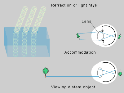 |
| Light rays bend as they pass through
water, glass, crystal, plastic or other
transparent material. This is called refraction.
Light rays from various distances bend to a
different angle as they pass through the lens of
the eye. The lens accommodates by adjusting in
thickness to place the light rays on the retina. |
Then, using special charts with letters and numbers,
the doctor is able to detect whether images are in
focus. For preschoolers who cannot read, the eye exam is
performed using illustrations instead of letters and
numbers.
The characters are scaled in size as if they were 3/8
inch high when viewed from 20 feet. "Normal" vision can
distinguish letters this size at this distance. This is
referred to as 20/20 vision.
To determine the factors causing any focusing errors,
the eye doctor positions a large instrument in front of
your eyes. You read the eye chart while the eye doctor
rotates lenses in front of your eyes, and you provide
feedback regarding your vision. Once your optimum vision
is reached, the eye doctor records the strength of the
lenses that provided this clarity.
You should feel free to ask your eye doctor what each
test measures, what your results are, and about the
latest equipment and technology.
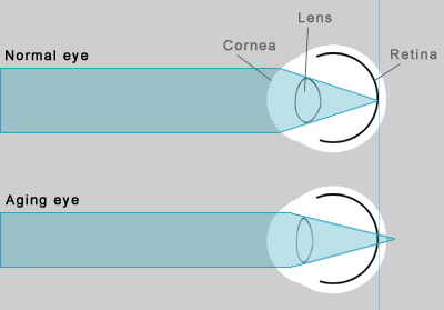 |
| A nearsighted eye is too long, causing
light to converge in front of the retina and
blurring distant vision. |
Retinal Exam
During a retinal exam, or ophthalmoscopy, the eye doctor
views the back of your eye through an instrument called
an ophthalmoscope. The retina should be examined
regularly so that problems can be discovered before they
damage vision. There are other tests that would be
performed if the ophthalmoscopy indicates potential
problems.
The retina is best examined through a dilated pupil,
allowing the eye doctor a wider view inside the eye. Eye
drops are used to dilate the pupil. The drops cause
temporary blurriness and sensitivity to light but these
side effects subside relatively quickly.
Prior to your exam, you should share your vital
health information with your eye doctor, and parents
should do so for their children. If you change eye
doctors, try to provide your new doctor with your prior
records. Be sure to discuss:
- medications you are taking
- allergies
- health conditions such as diabetes
- recent illness
Routine exams are varied somewhat based on age and
eye condition.
Birth
At birth, a baby's first medical examination includes a
check for congenital eye defects. Although rare, early
diagnosis of these problems is important to preserve
sight. Pediatricians and eye doctors can usually correct
most eye problems, if spotted early.
Some babies are born with strabismus, in which one or
both eyes are not straight. It can be caused by either
extreme farsightedness or eye muscle imbalance. The
condition usually goes away within a few months without
treatment. Premature babies have a greater chance of
developing this condition.
Strabismus can develop from birth to about age 7. It
may be rarely noticed at first but become frequent over
time.
Infancy
At 3 to 4 months, parents should consult an eye doctor
if a baby:
- cannot focus on or follow an object with both
eyes
- has difficulty moving one or both eyes in all
directions
- has crossed eyes most of the time
- has one or both eyes that tend to wander outward
Otherwise, infants should have an eye exam at the age
of 6 months. During a thorough examination, the eye
doctor will test both of the baby's eyes for large
differences in visual status to rule out "lazy eye," or
amblyopia. He or she will check the baby's eye movements
and eye health. Problems with vision development and eye
health are uncommon in infants but most easily treated
if caught early.
Childhood and Adolescence
Normally, parents should schedule an eye examination
when their child is 3 years old, then again just before
school begins. Once of school age, regular exams are
recommended once a year even if no problems have been
detected previously. Visual changes take place gradually
and may go unnoticed by a child, parents, and
caretakers.
Visual screenings done in school are valuable for
spotting conditions that could affect how a child is
functioning. These are preliminary assessments, designed
to detect vision defects such as nearsightedness and
farsightedness, and give a rough degree of refraction
error. The visual screenings are no substitute for a
more thorough evaluation by an eye doctor. It is
estimated that 25 percent of school-age children with
correctable vision problems do not receive treatment.
- Eye Conditions - Most preschool and young
school-aged children are slightly farsighted. The
condition lessens as children grow, usually
stabilizing by adolescence. Nearsightedness,
however, may begin in childhood but continue to
progress through adolescence and into early
adulthood. Parents' first clues that their child is
nearsighted often occur at school. When a child has
difficulty seeing the blackboard, learning or
behavioral problems often surface in class. Because
of vision difficulties, the child may avoid sports.
Other clues are:
- sitting too close to the television
- eye rubbing
- squinting
- clumsiness
- holding head at an odd angle
- headaches or dizziness
During childhood vision changes quickly.
Experts recommend eye exams every six months for
children who need glasses.
- Injury and Infection - The most common
need for eyecare in childhood is caused by infection
and injury.
Pink eye (conjunctivitis), corneal scratches and
sports injuries are the most common causes.
For a child of any age, parents should consult an
eye doctor if they notice any of the following,
which could indicate injury of infection:
- an eye that wanders inward or outward when
tired
- eyelid droopiness
- redness of eyes or eyelids
- crusted eyelids
- tearing or leaky eyes
- eyelid styes or sores
- too much eye rubbing
- avoidance of bright light
Adulthood
The human eye reaches peak strength in the young adult,
around the mid- to late-20s. Night vision, eye-hand
coordination, motion and depth perception, and color
discrimination may all improve during this time. During
the years of improved sight, nutrition can contribute to
optimum vision.
Aging Eyes
By the time we reach our mid- to late-30s, most people
begin to have difficulty focusing on close objects. The
ciliary muscles that adjust the thickness of the lens
start to weaken. Meanwhile, the lens itself loses its
elasticity. Consequently, the ability of the lens to
focus at close range decreases. The condition is
referred to as "aging eyes" or presbyopia.
Most people notice signs of aging eyes, between the
ages of 40 and 45 when they begin holding reading
material at arms' length. The majority need to wear
reading glasses or other corrective lenses. The
condition may progress indefinitely or stabilize by 65
to 70 years of age. To detect this condition, an annual
eye exam is recommended.
Sensitivity to Light
During our mid-40s, the iris muscles tend to slow. The
reflex response decreases, increasing the amount of
light entering the eye. For many people, sensitivity to
glare starts at this age. Their eyes may be overexposed
to the sun and other UV light, potentially causing
permanent damage. Eyewear with ultraviolet radiation
protection can shield the eyes from the harmful rays.
Pregnancy
Vision often changes during pregnancy but usually
returns to normal after delivery. Here are some
eye-related changes that may occur during pregnancy:
- a change in refraction, requiring a different
prescription for corrective eyewear
- blurry vision
- dry eye
- less tolerance of contact lenses
- worsening of existing eye conditions
Vision problems during pregnancy may signal other
health problems. Blurred vision or seeing spots may
indicate gestational diabetes or pregnancy-induced
hypertension, an increase in blood pressure that usually
occurs after the 20th week of pregnancy. Eclampsia and
pre-eclampsia, caused by extremely high blood pressure,
can cause eye hemorrhages and retinal detachment,
although these are extremely rare.
Not all pregnant women develop eye problems, but
experts recommend routine examinations by an eye doctor
each trimester. Early treatment is vital to the health
of the mother and baby.
Maturity
Healthy eyes and good vision help older people lead
active, independent lifestyles. Many eye problems are
treatable, especially if spotted early. An annual visit
to the eye doctor is one of the best ways to maintain
healthy eyesight. This is especially important if you
have diabetes or eye disease.
Some vision changes are a normal part of aging.
Others may be indicative of other diseases such as high
blood pressure or diabetes, both common in older adults.
Common occurrences during the elder years include:
- stabilization of presbyopia, or "long arm
syndrome"
- development of cataracts
- increase in spots, floaters and flashes
- onset of glaucoma
- incidence of dry eye
- disease, infection or injury of the cornea
- retinal disorders such as macular degeneration,
diabetic retinopathy and retinal detachment
Retinal disorders are a leading cause of vision loss
in elderly people. When damaged, the light-sensitive
cells lining the retina cannot pass images to the brain.
If detected and treated early, vision loss may be slowed
or halted.
Retinal Detachment
During the aging process, the retina may become detached
from the back of the eye. If caught in time, laser
surgery may be able to bond it to the back of the eye.
You should consult an eye doctor if you experience:
- blurry central vision
- clouded vision
- inability to see faces or details clearly
- double vision
- visual distortion
- sudden vision loss
- sudden onset of flashing lights
Corneal Disease
Older adults are particularly vulnerable to corneal
disease, largely due to the high incidence of dry eye.
Symptoms include:
- redness
- reflex tearing or watery eyes
- eye pain
- loss of vision
- seeing halos
Accommodation
In addition to corrective eyewear and regular eyecare
exams, you can make minor changes in your home or in
your behavior to adapt to the limitations of your
changing vision. There are several ways that you can
enhance the safety and convenience of your surroundings:
- Make sure lighting is adequate, day and night,
in key areas:
- outdoors
- garage
- storage areas
- stairways
- work spaces
- favorite reading places
- Keep flashlights in your car and in a carryall
bag for unexpected situations.
- If your vision is best in the morning, schedule
your reading and detailed work accordingly.
- If your ability to see in the dark is diminished
or you have increased sensitivity to glare:
- Use caution when walking near traffic.
- Drive only on well-lit roads.
- Keep your windshield, headlights and glasses
clean.
- Wear anti-glare glasses.
- Consider avoiding driving at night
Sources
Cassel, G. Billig. The Eye Book: A Complete Guide to
Eye Disorders and Health. Baltimore, MA: Johns
Hopkins University Press, 1988.
Collins, J.F. Your Eyes: An Owner's Guide.
Englewood Cliffs, NJ: Prentice Hall, 1995.
D'Alonzo, T.L. Your Eyes: A comprehensive Look at the
Understanding and Treatment of Vision Problems.
Clifton Heights, PA: Avanti Publishing, 1991.
Eden, J. The Physician's Guide to Cataracts,
Glaucoma, and Other Eye Problems. New York, NY:
Consumer Reports Books, A Division of Consumers Union
Yonkers, 1992.
Schuman, B.N. The Human Eye. New York, NY:
Atheneum, 1986.
Leach, Penelope. Your Baby and Child. Alfred A.
Knopf. New York, NY: 1990
Benjamin, William J, ed. Borish's Clinical Refraction.
Montreal, Canada: W.B. Saunders, 1998. |
