 |
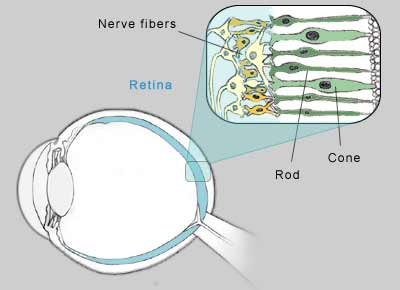
| Rods are responsible for night peripheral
vision. Cones produce color and central vision. |
The wide, cone-shaped cells of the retina are
sensitive to light. In bright environments, the chemical
iodopsin increases in the cones. They become more
sensitive, and we can see greater detail and color.
The long, thin rods react to lack of light. In the
dark, a chemical called rhodopsin, or visual purple,
increases in the rods, improving their sensitivity.
Meanwhile, the cones do not receive enough light for a
chemical reaction to take place, so they do not function
in the dark. This provides vision in dim light, but with
less detail.
The rods require about 30 minutes to be fully
functional in the dark, while the cones adapt to
brighter conditions in just a few minutes.
Central and Peripheral Vision
The light-sensitive rods and cones form a tightly packed
network.Each eye contains 100 million rods and 3 million
cones but they are not distributed evenly. Rods are
mainly located in the outer edges of the retina, and
cones are clustered in the center. As a result, central
vision is clearest, while peripheral vision is less
precise.
Vision Persistence
The image of an object on the retina is retained very
briefly, fading almost instantly. But the eyes
instinctively continue to gather new input. In a healthy
retina, new images are received before old images fade -
at the rate of about 30 times per second. This gives the
appearance of an image merging into the next. It is this
persistence of vision that provides the appearance of
continuous, smooth movement - just as motion pictures
blend one frame into another.
Binocular and Stereoscopic Vision
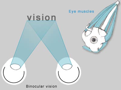 |
| Muscles coordinate the position and
movement of each eye, creatin binoular vision. |
"Binocular vision" refers to sight generated from two
eyes. The separate images received in each eye are
combined into a single image. To see clearly, without
double vision, the images must fall precisely on
corresponding positions on each retina. Six muscles
surrounding each eye work together to position the eyes
properly so that light is focused on the center of each
eye, providing clear vision. To focus on objects that
are within close range, the eyes move closer together
(convergence). To focus further away, the eyes move
further apart (divergence). When the eyes are not
aligned properly, double- vision results. Binocular
vision is largely responsible for our ability to see in
three dimensions. The slight difference in the angles of
the images received in each eye gives images depth. This
ability is called stereoscopic vision.
However, people with vision in only one eye do not
necessarily see flat, two-dimensional images. Light,
shade, shadows, color and relative sizes of objects
contribute to depth perception.
People think and learn best in three dimensions. When
scanning text quickly, we can absorb 100 letters per
second - the computer equivalent of 100 bits per second.
By comparison, when glancing at a three-dimensional
object, we can see the equivalent of 1 billion bits per
second.
Eye-Brain Connection
Visual information is received through the eyes but
interpreted with the brain. Electrical signals are
relayed from the retina to the brain via the optic
nerve. The ability to recognize what we see lies in the
occipital lobe, near the back of the head.
The Blind Spot
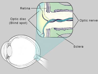 |
| The area where the optic nerve meets the
retina is a "blind spot". |
In the small, round area where the optic nerve meets
the retina, there are no rods or cones. If light from an
object lands on this spot, the image is essentially
invisible. This area is called the optic disk, or the
blind spot. In effect, it's a black hole in the retina's
projection screen.
This missing area in the field of vision is not
normally noticeable. The brain fills this blind spot
with the colors and patterns of the objects surrounding
environment. It uses information received fractions of a
second prior from a different distance or angle and what
the opposite eye sees to create a complete,
uninterrupted picture.
Everyone has this blind spot in each eye. The size of
the blind spot varies from one person to another, and
even from one eye to the other. To observe the effects
of the blind spot, use the illustration below:
 |
| Images "disappear" when projected on the
blind spot. The brain fills in the "hole" with
details from the background. |
Close your left eye. Look at the dot with your right
eye from about one foot away, then gradually move toward
it. Be sure to keep your eye on the dot and move very
slowly. At some point, the dot will disappear. That is
the distance and angle at which the dot is reflected on
the optic disk.
Notice that the brain fills in the blind spot. It
guesses what's in the area that it can't see. In this
case, it fills it with black, like the area immediately
around the dot.
Similarly, if you were to draw a line across the
screen and through the dot, and repeat the exercise, the
dot would disappear but your brain would fill in the gap
in the line.
Optical Illusions
Sometimes the brain distorts reality, incorrectly
interpreting the environmental clues that surround an
object. Optical illusions demonstrate this phenomenon.
Which person is tallest?
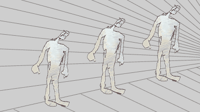 |
| Which figures are tallest and smallest? |
From an early age, people learn that the more distant
an object is, the smaller it looks. In the illustration
above, the person on the far right appears to be smaller
than the person on the left. They are actually the same
size.
People who live in the jungle often have trouble
judging distances in the open country. Because their
environment has not contained open, distant views, they
have not learned that distant objects look smaller.
What do you see?
 |
| Do you see vase or two faces? |
Sometimes there is more than one way for the brain to
interpret an image. In the illustration above, do you
see a vase or two faces? The vase is white on a black
background, while the faces are black on a white
background.
Which square is larger?
 |
| Eye-Brain Communication Fig 6 |
In each of the two illustrations above, one square is
surrounded by four rectangles larger than the center
square in one drawing, smaller in the other. The center
square that is surrounded by smaller rectangles appears
to be larger than the center square surrounded by larger
rectangles. In reality, the center squares are the same
size.
The Moon
Why does the moon appear larger when it is near the
horizon than it does overhead?
When the moon is close to the horizon, it is viewed
along with other objects, such as mountains or
skyscrapers. Without the benefit of nearby objects, the
moon seems to shrink in the expansive sky. The brain
misinterprets the size of the moon relative to its
surroundings.
Virtual Sight
Experiments have revealed that by stimulating the retina
with preprogrammed configurations of ultrasound, some
blind people are able to "see" black and white outlines
and images. The brain interprets the images as if light
were projected on the retina.
If a camera could successfully digitize images and
translate them into recognizable ultrasound impulses, it
may be possible to give virtual sight to the blind.
Sources
Cassel, G. Billig. The Eye Book: A Complete Guide to
Eye Disorders and Health. Baltimore, MA: Johns
Hopkins University Press, 1988.
Collins, J.F. Your Eyes: An Owner's Guide.
Englewood Cliffs, NJ: Prentice Hall, 1995.
D'Alonzo, T.L. Your Eyes: A Comprehensive Look at the
Understanding and Treatment of Vision Problems.
Clifton Heights, PA: Avanti Publishing, 1991.
Eden, J. The Physician's Guide to Cataracts,
Glaucoma, and Other Eye Problems. New York, NY:
Consumer Reports Books, A Division of Consumers Union
Yonkers, 1992.
Schuman, B.N. The Human Eye. New York, NY:
Atheneum, 1986.
Adler, R., Adler, I. Your Eyes. New York, NY: The
John Day Company, 1992.
Begbie, G.H. Seeing and the Eye: An Introduction to
Vision. Garden City, NY: National History Press,
1996.
Cohen, N.S. Out of Sight Into Vision: There is More
to Good Vision Than Reading the Fine Print. Toronto,
Canada: Collier Macmillan Canada, 1997.
Kwiko, M.L. Eyes. Toronto, Canada: Key Porter
Books, 1994.
Rainwater, J. Vision: How, Why, and What We See.
New York, NY: Golden Press, 1992.
Leach, Penelope. Your Baby and Child. Alfred A.
Knopf. New York, NY: 1990
Benjamin, William J, ed. Borish's Clinical Refraction.
Montreal, Canada: W.B. Saunders, 1998. |
